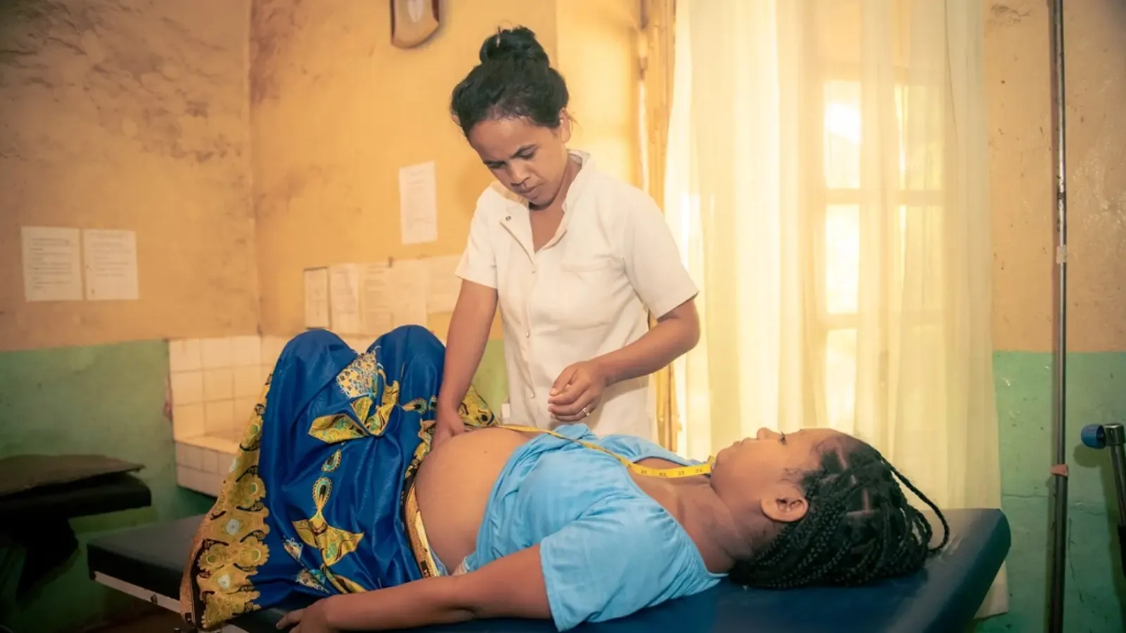
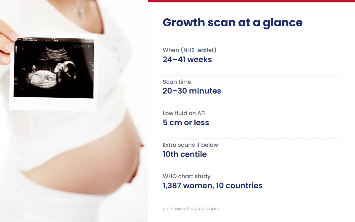
A growth scan is a third-trimester ultrasound that checks how the baby is growing. It measures the head, abdomen and thigh bone, estimates the weight, and checks the fluid and the blood flow in the cord. One NHS hospital leaflet says it can be done at any time from 24 to 41 weeks. The weight it gives is an estimate with a known margin of error, and your doctor reads it with your own history.
What Is a Growth Scan?
A growth scan in pregnancy uses the same ultrasound probe as earlier scans, moved over the bump with gel. The Gateshead NHS leaflet says it takes about 20 to 30 minutes. The sonographer measures the baby, the machine turns those measurements into an estimated fetal weight, and the result is plotted on a chart for the week of pregnancy.
It is different from the two scans most women have earlier. The NHS offers a dating scan at 11 to 14 weeks and a screening scan at 18 to 21 weeks. This third-trimester scan comes later and follows size and wellbeing over time.
In India, the Ministry of Health and Family Welfare runs the Pradhan Mantri Surakshit Matritva Abhiyan, which offers free antenatal check-ups on the 9th of every month. Its minimum package includes one ultrasound during the second trimester. The World Health Organization recommends one ultrasound before 24 weeks for every pregnant woman, to date the pregnancy and detect anomalies and twins.
A late scan is not part of routine care for every pregnancy. A Cochrane review of 13 trials with 34,980 women found that routine ultrasound after 24 weeks in low-risk or unselected pregnancies did not benefit mother or baby. That is why a pregnancy growth scan is usually ordered for a reason.
When and Why the Scan Is Done
The Gateshead leaflet says women judged moderate or high risk may have a series of scans in the third trimester, booked one at a time by the doctor. It lists reasons for an urgent scan:
- repeated episodes of reduced baby movements
- a medical condition diagnosed during the pregnancy
- a bump that measures small, or grows slowly, when the midwife checks the fundal height
- slowing growth on an earlier scan
Growth scan weeks are therefore set by your own pregnancy. A 32 weeks pregnant scan report uses the same terms as one at 36 weeks; only the expected values change with the week. Follow your hospital's own advice on preparation, such as whether to come with a full bladder.
What a Growth Scan Measures
Fetal biometry is the set of body measurements taken on the screen. The table lists the terms you will see on the report and what each one is for.
| Report term | What it measures | How it is used |
|---|---|---|
| BPD and HC | Width of the head, and distance around it | Head size for the week |
| AC | Distance around the abdomen | Body size, read with the head |
| FL | Length of the femur, the thigh bone | Bone growth for the week |
| EFW | Weight calculated from head, abdomen and femur | Plotted as a centile on a growth chart |
| AFI or SDP (liquor) | Amniotic fluid around the baby, in cm | 5 cm or less on the AFI suggests low fluid |
| Umbilical artery Doppler (PI) | Blood flow from the placenta along the cord | Raised PI means the placenta is working harder |
How to Read a Growth Scan Report
Size and centiles
A centile places the baby among 100 babies at the same week of pregnancy. On the 50th centile, half are bigger and half smaller; on the 10th, about 10 in 100 are smaller. The Gateshead leaflet arranges extra scans when growth slows or the baby measures below the 10th centile or above the 97th. Hospitals use different charts, so the same EFW can sit on a slightly different centile from one report to another.
Fluid terms
Liquor is the report's word for amniotic fluid. The amniotic fluid index (AFI) adds the deepest pocket of fluid in each of four quadrants of the womb, and the single deepest pocket (SDP) uses one pocket. A Cochrane review describes an AFI of 5 cm or less, or no pocket measuring 2 × 1 cm, as a sign of reduced fluid. In 4 trials with 3,125 women, the AFI labelled low fluid 2.33 times as often as the single pocket and led to more inductions, with no better outcome for babies. Too much fluid, called polyhydramnios, also gets a closer look.
Doppler terms
Doppler measures blood flow. The umbilical artery result is usually given as a pulsatility index (PI), and the Gateshead leaflet explains a raised PI as the placenta working harder to push blood along the cord. Some reports add the middle cerebral artery, a vessel in the baby's brain. A Cochrane review of 19 trials with 10,667 women found that umbilical artery Doppler in high-risk pregnancies was linked with fewer perinatal deaths, 1.2% against 1.7%.
Position and placenta terms
Cephalic means head down, breech means bottom or feet first, and transverse means lying across the womb. The placenta is described by where it sits, such as anterior (front wall), posterior (back wall) or fundal (top). A placenta reported as low-lying is followed up by your doctor. The fetal heart rate appears in beats per minute.
How Accurate Is the Estimated Fetal Weight?
The weight on the report comes from a formula, so it carries the error of every measurement fed into it. A Bengaluru study in the Journal of Medical Ultrasound tested 34 published formulas on 149 Indian pregnancies. Only 12 had an average error within ±10%, and of those, only four placed more than 60% of estimates within 10% of the actual birth weight. The authors measured an average random error of 10.41% and traced it to differences between operators, their training, image quality and machine calibration.
Charts matter too. The WHO fetal growth charts were built from 1,387 low-risk pregnancies in 10 countries, India among them. That study found Indian newborns were significantly smaller than those in the other countries, even after adjusting for weeks of pregnancy. It also found that each extra 10 cm of the mother's height added about 1% to 2% to the estimated weight.
So one EFW in pregnancy is a snapshot. Two scans a few weeks apart show whether the baby is growing steadily, which is what the doctor watches. For average weights week by week, see the pregnancy weight chart for mother and baby.
What the Scan Cannot Tell You
Finding the sex of the foetus by scan is illegal in India: the PCPNDT Act prohibits sex selection and regulates prenatal tests so they are not misused to detect sex.
A scan also cannot judge the baby's wellbeing on its own. The Gateshead leaflet says a scan reassures only for the moment it is done, and reduced movements still need a call to the hospital, even on a day you had a scan. Ultrasound itself is considered safe: the NHS says there are no known risks to the baby or the mother from an ultrasound scan.
This is general information. Ask your doctor about your own readings.
Weighing Yourself at Home During Pregnancy
An ultrasound follows the baby, and a scale at home follows the mother. Weigh at the same time of day, on the same hard floor and in similar clothes, and note the reading for your next antenatal visit. A home reading is information for your doctor, never a target.
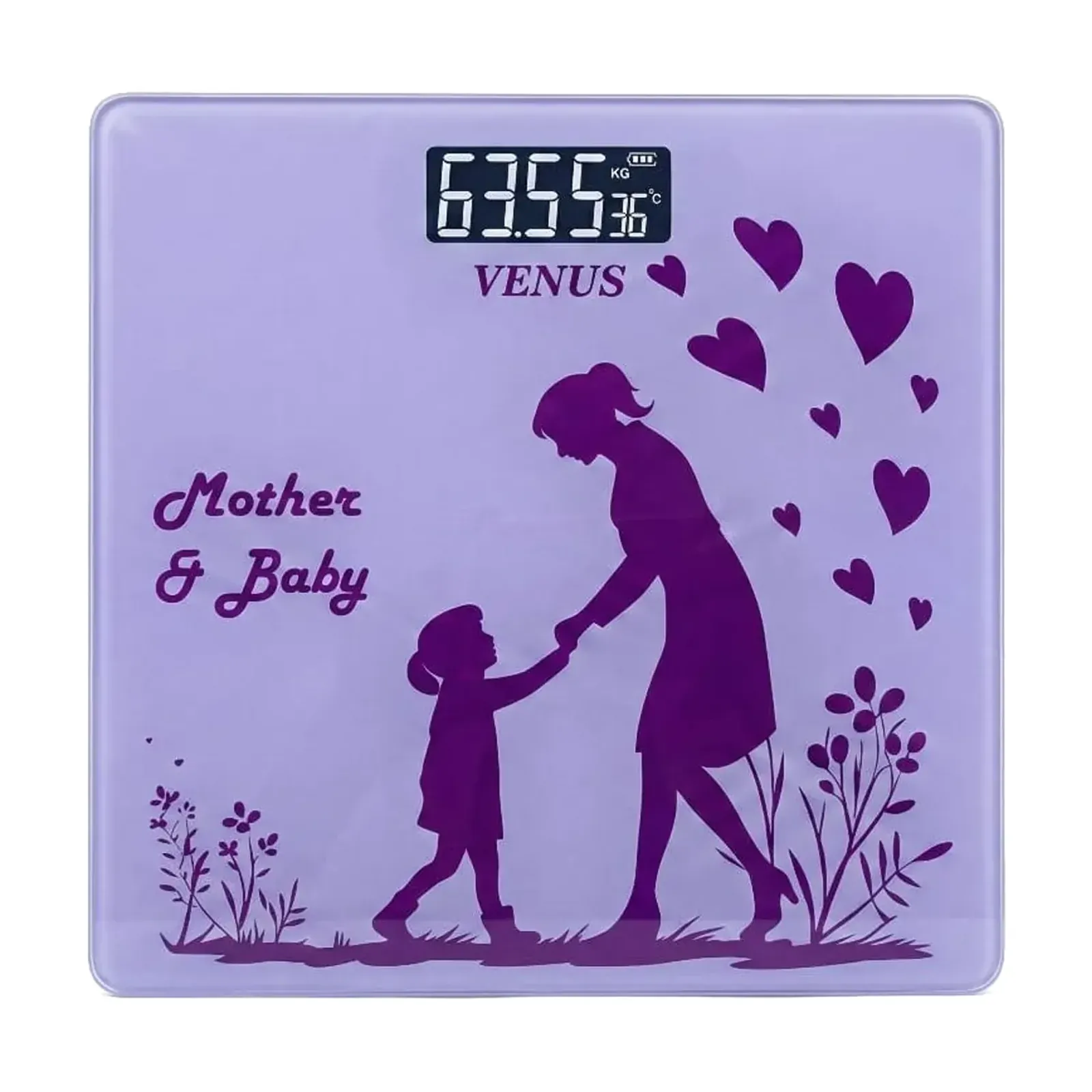
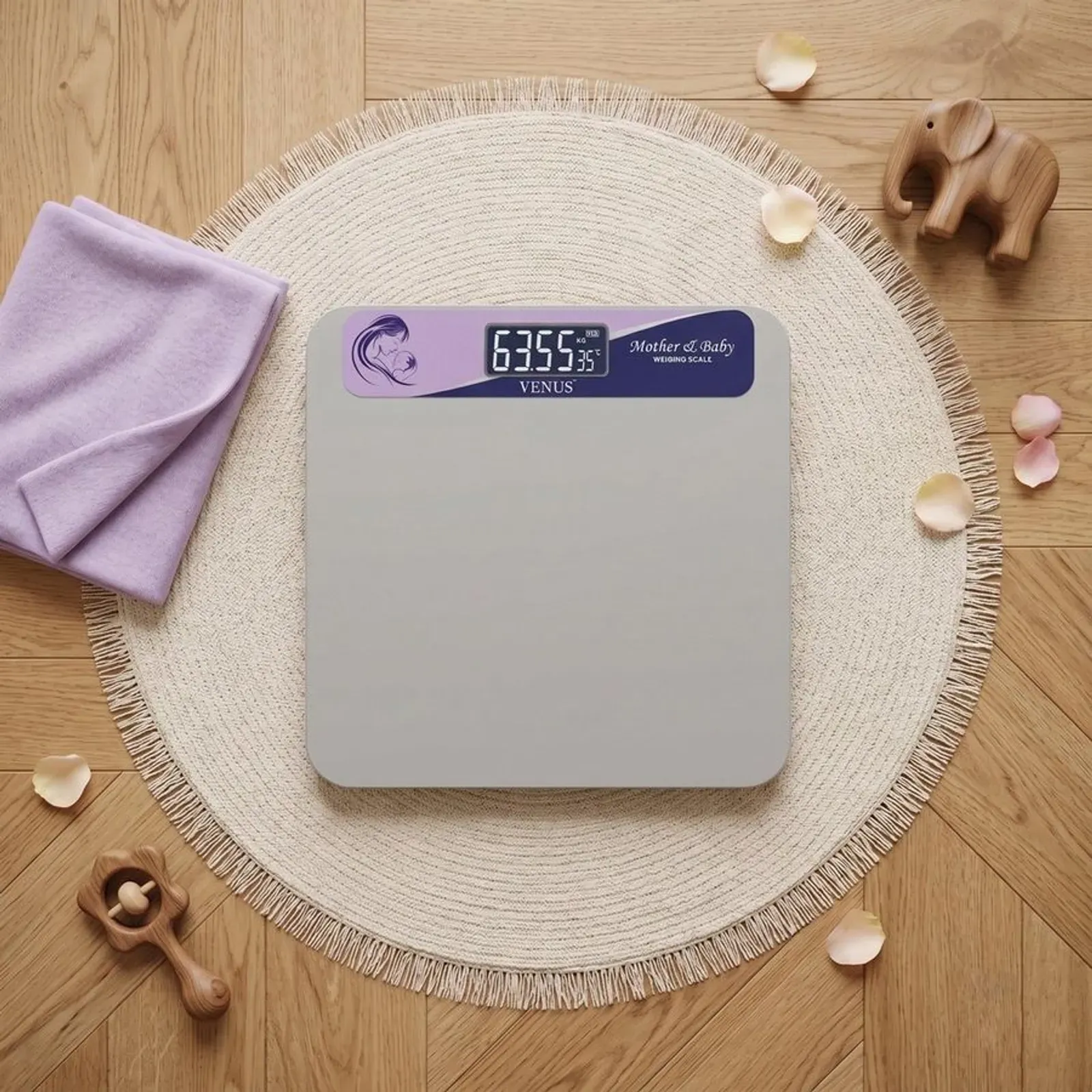
The Venus EPS-05 mother and child weighing scale weighs up to 180 kg on a 28 × 28 cm glass platform and costs ₹729. After birth, its auto tare mode gives the baby's weight while the mother holds the baby on the scale. The Venus EPS-09 (M) mother and baby weight machine has the same auto tare and a steel top, also for up to 180 kg, at ₹1,040.
Why Buy From a Certified Indian Manufacturer
Omace Brands Private Limited has made weighing scales in Bagru, Jaipur since 1994, as the About Omace page records. As a manufacturer, the company holds ISO 13485:2016 and EU MDR quality certificates, both written for medical device makers. It also holds ISO 9001:2015, a CE certificate of compliance and a US FDA compliance certificate, all listed on the certificates page. Every scale is checked with certified test weights before dispatch. Every Omace digital scale opens calibration mode when TARE is held for about 5 seconds. Scales from the Omace store carry a 2-year warranty on manufacturing defects, and every Omace product is made in India.
Before Your Next Antenatal Visit
Keep each scan report in one folder, in date order, so the doctor can compare the EFW, its centile, the fluid and any Doppler result across visits. Ask whether the size fits your weeks, whether growth has been steady since the last scan, and when the next check is due. After birth, the baby's own weight becomes the record; the newborn weight chart explains the first months, and the baby weighing scales range covers home and clinic use.